
Medical Imaging and Image Processing
Medical care goes hand in hand with analysing and using a very large and constantly growing amount of medical data. Imaging diagnostics, for example, is one of the two main pillars in the detection and diagnosis of diseases. The task of radiologists is initially to create medical images using MRI, CT and co. The images are then either examined for abnormalities (detection) or the presenting clinical picture is characterised (diagnosis), depending on the issue at hand. This diagnostic process is time-consuming and tedious in view of the large volumes of data involved. Very time-consuming and complex image processing procedures are also often necessary for the planning and management of minimally invasive interventions in therapy.
The research field of medical image processing has therefore been dominated for several years by machine learning topics, especially with the help of deep neural networks (deep learning).
AI-Safety-Monitoring
Contact: Prof. Dr. Stefanie Remmele, Prof. Dr. Eduard Kromer
Duration: February 2026 – May 2028 (2.5 years)
Funding: BMWi, ZIM
Partners: deepc GmbH, LMU Department of Radiology
Research Associate: Tobias Ziegler
A key challenge in the use of AI systems in medical imaging is what are known as “domain shifts.” This term refers to discrepancies between the data used to train algorithms and the real-world data encountered in everyday clinical practice.
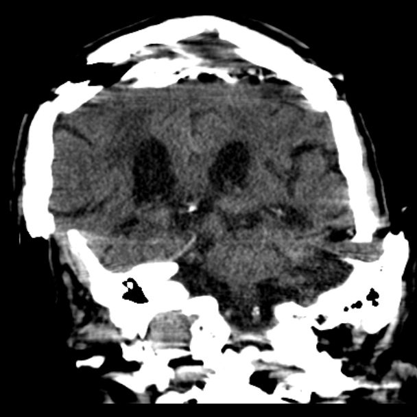
Such differences can arise, for example, from variations in CT scanners or examination protocols. Differences in patient groups or image artifacts (Image source: Case courtesy of David Puyó Vera, Radiopaedia.org, rID: 25637) resulting from motion or foreign material can also affect the reliability and performance of AI models. This is precisely where the AI Safety Monitoring project comes in. The goal is to detect domain shifts early on in order to sustainably increase the transparency and trustworthiness of AI models in medical imaging.
Caption: CT image with motion artifact (Case courtesy of David Puyó Vera, Radiopaedia.org, rID: 25637)
NeuroTEST
Contact: Prof Dr Stefanie Remmele
Duration: 2021-2023 (2 years)
Funding: ZIM
Partner: deepc GmbH, Munich
Associated partners: LAKUMED Krankenhaus Landshut-Achdorf, Prof Dr Tobias Schäffter (PTB Berlin, TU Berlin)
Research assistant: Christiane Posselt
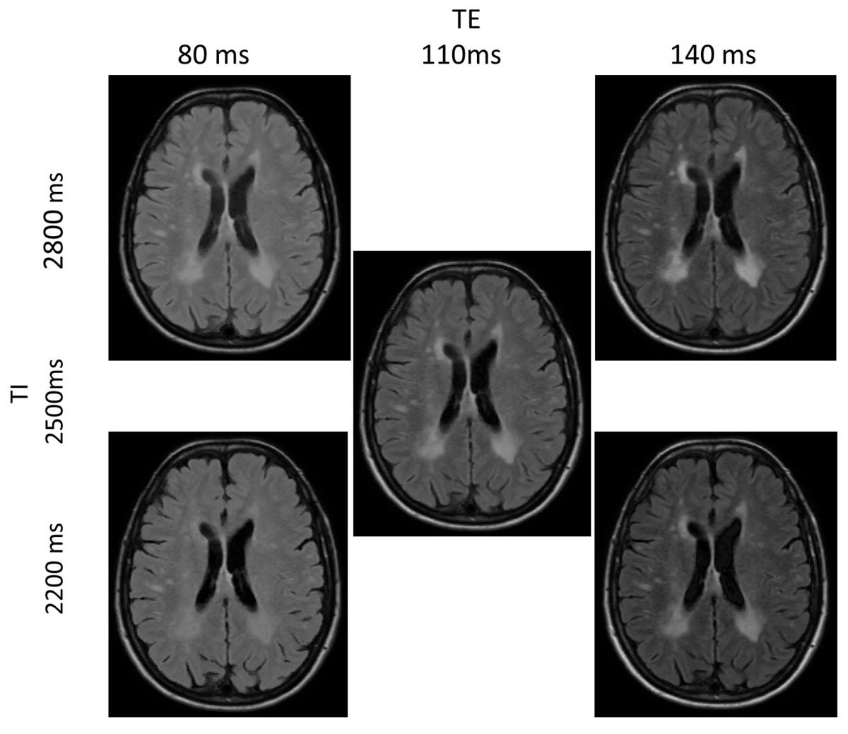
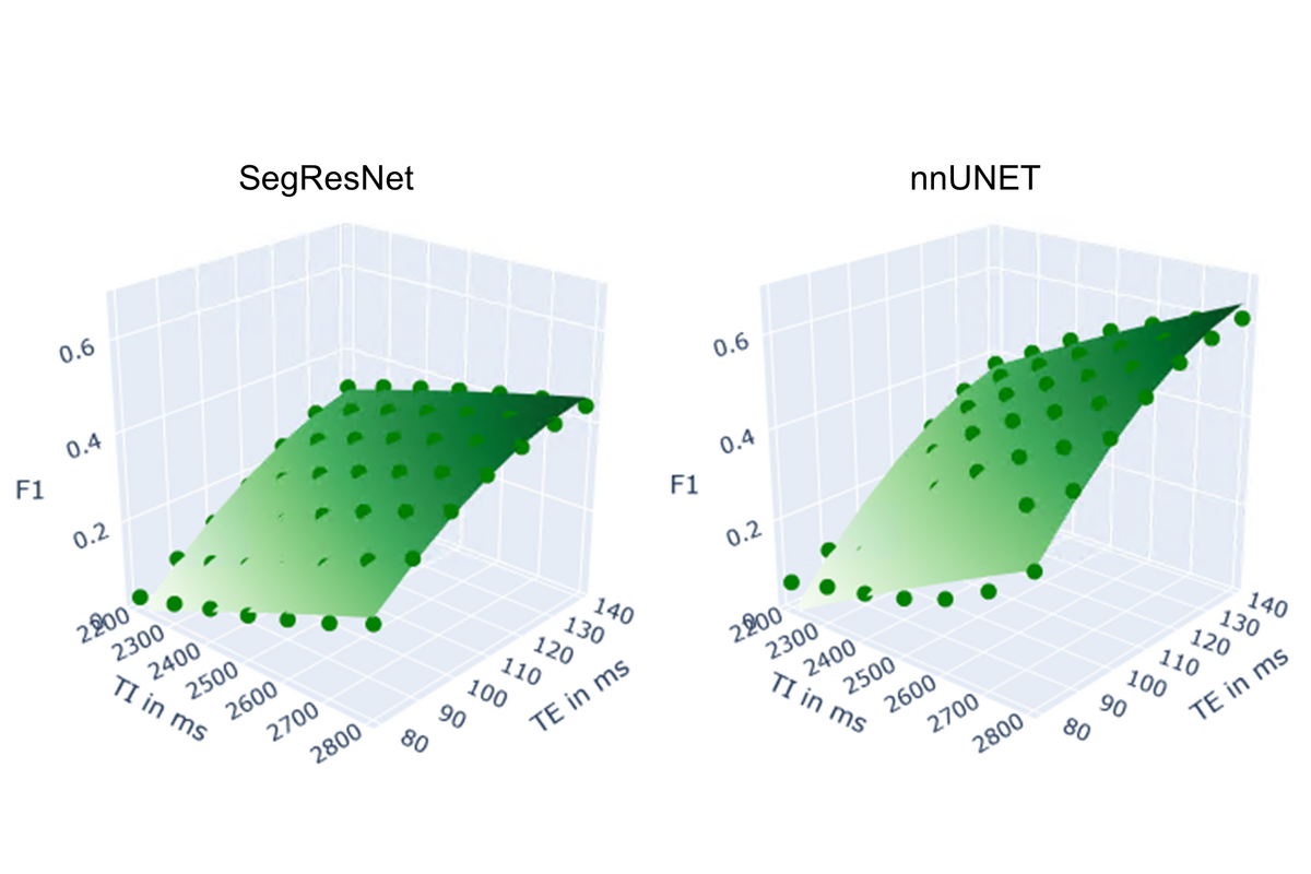
Machine learning methods, in particular using deep neural networks, are celebrating ever new successes in the analysis and classification of medical images. It is clear that in many applications, the algorithms are already achieving at least human-like decision-making accuracy. However, it is still unclear how networks trained on a specific data situation will behave in a different data situation. For example, when imaging hardware or imaging parameters differ from radiology to radiology.
In the BMWi-funded ZIM project NeuroTEST, the Medical Technology research group (project lead Prof Remmele) is developing methods for the systematic validation of neural networks (use case: segmentation of MS lesions) together with the Munich start-up deepc. To this end, methods of statistical experimental design are being researched, whereby AI algorithms are systematically tested on data from different recording protocols. The project at HAW focuses on the simulation and synthesis of this data in order to be able to provide arbitrary data domains for the stress tests.